




Показаны сообщения с ярлыком днк. Показать все сообщения
Показаны сообщения с ярлыком днк. Показать все сообщения
17.02.2013

18.05.2012
Чем грозит устранение бесплодия будущим эмбрионам?
Наткнулась на статью на
http://www.laboratoryequipment.com/news-Fertility-Treatment-Heightens-Risk-of-Birth-Defects-050712.aspx о том, что применение некоторых средств борьбы с бесплодием повышает риск появления дефектов развития у плода... Интересное исследование...
Fertility Treatments Heighten Risk of Birth Defects
May 7, 2012

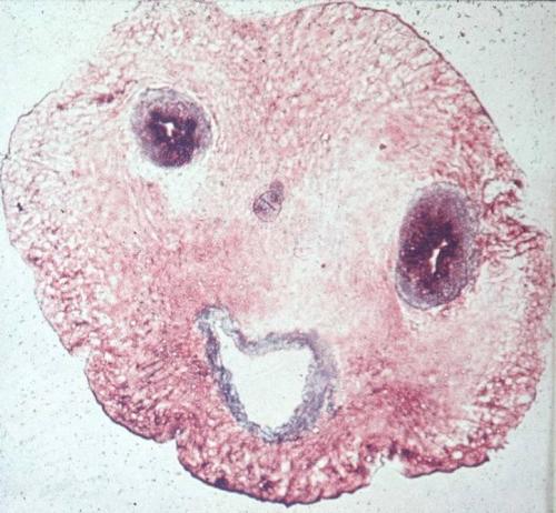
A six week embryonic age or eight week gestational age intact Embryo, found in a Ruptured Ectopic pregnancy case. Image: Vilas Gayakwad, Wikimedia
A Univ. of Adelaide study has identified the risk of major birth defects associated with different types of assisted reproductive technology.
In the most comprehensive study of its kind in the world, researchers from the Univ.’s Robinson Institute have compared the risk of major birth defects for each of the reproductive therapies commonly available internationally, such as: IVF (in vitro fertilization), ICSI (intracytoplasmic sperm injection) and ovulation induction. They also compared the risk of birth defects after fresh and frozen embryo transfer.
The results are being published in the prestigious New England Journal of Medicine, and presented in Barcelona, Spain at the World Congress on Building Consensus in Gynecology, Infertility and Perinatology.
"While assisted reproductive technologies are associated with an increased risk of major birth defects overall, we found significant differences in risk between available treatments," says the lead author of the study, Associate Prof. Michael Davies from the Univ. of Adelaide’s Robinson Institute and School of Paediatrics and Reproductive Health.
Researchers linked a census of more than 6100 assisted reproductive technology births in South Australia to a registry of more than 300,000 births and 18,000 birth defects. They compared risks of birth defects across all infertility treatments to pregnancies in women with no record of infertility. They also compared successive pregnancies for women.
Previous studies have identified an increased risk of birth defects associated with infertility treatment, but this is the first study to compare all forms of available treatment. This is also the first study to compare pregnancies within women by the treatments received.
"The unadjusted risk of any birth defect in pregnancies involving assisted conception was 8.3 percent (513 defects), compared with 5.8 percent for pregnancies not involving assisted conception (17,546 defects)," Davies says.
"The risk of birth defects for IVF was 7.2 percent (165 birth defects); and the rate for ICSI was higher at 9.9 percent (139 defects). A history of infertility, either with or without assisted conception, was also significantly associated with birth defects. While factors associated with the causes of infertility explained the excess risk associated with IVF, the increased risk for a number of other treatments could not readily be explained by patient factors. ICSI, for instance, had a 57 percent increase in the odds of major defect, although the absolute size of the risk remained relatively small," he says.
Davies says cryopreservation (freezing) of embryos was associated with a substantially reduced risk of birth defects, particularly for ICSI. "This may be due to developmentally compromised embryos failing to survive the freeze/thaw process," he says.
Also of concern was the tripling of risk in women using clomiphene citrate to stimulate ovulation outside of a closely supervised clinical setting.
"While confined to a small group in our study, this is of particular concern as clomiphene citrate is now very widely available at low cost, and may easily be used contrary to manufacturers' very specific instructions to avoid use if pregnant, as it may cause fetal malformations. This aspect of the study will need additional confirmation from future research," Davies says.
He says the study now needs to be expanded to include more recent years of treatment, as the reproductive technologies have undergone continual innovation which may influence the associated risks of treatment.
Source: Univ. of Adelaide
Fertility Treatments Heighten Risk of Birth Defects
May 7, 2012
A six week embryonic age or eight week gestational age intact Embryo, found in a Ruptured Ectopic pregnancy case. Image: Vilas Gayakwad, Wikimedia
A Univ. of Adelaide study has identified the risk of major birth defects associated with different types of assisted reproductive technology.
In the most comprehensive study of its kind in the world, researchers from the Univ.’s Robinson Institute have compared the risk of major birth defects for each of the reproductive therapies commonly available internationally, such as: IVF (in vitro fertilization), ICSI (intracytoplasmic sperm injection) and ovulation induction. They also compared the risk of birth defects after fresh and frozen embryo transfer.
The results are being published in the prestigious New England Journal of Medicine, and presented in Barcelona, Spain at the World Congress on Building Consensus in Gynecology, Infertility and Perinatology.
"While assisted reproductive technologies are associated with an increased risk of major birth defects overall, we found significant differences in risk between available treatments," says the lead author of the study, Associate Prof. Michael Davies from the Univ. of Adelaide’s Robinson Institute and School of Paediatrics and Reproductive Health.
Researchers linked a census of more than 6100 assisted reproductive technology births in South Australia to a registry of more than 300,000 births and 18,000 birth defects. They compared risks of birth defects across all infertility treatments to pregnancies in women with no record of infertility. They also compared successive pregnancies for women.
Previous studies have identified an increased risk of birth defects associated with infertility treatment, but this is the first study to compare all forms of available treatment. This is also the first study to compare pregnancies within women by the treatments received.
"The unadjusted risk of any birth defect in pregnancies involving assisted conception was 8.3 percent (513 defects), compared with 5.8 percent for pregnancies not involving assisted conception (17,546 defects)," Davies says.
"The risk of birth defects for IVF was 7.2 percent (165 birth defects); and the rate for ICSI was higher at 9.9 percent (139 defects). A history of infertility, either with or without assisted conception, was also significantly associated with birth defects. While factors associated with the causes of infertility explained the excess risk associated with IVF, the increased risk for a number of other treatments could not readily be explained by patient factors. ICSI, for instance, had a 57 percent increase in the odds of major defect, although the absolute size of the risk remained relatively small," he says.
Davies says cryopreservation (freezing) of embryos was associated with a substantially reduced risk of birth defects, particularly for ICSI. "This may be due to developmentally compromised embryos failing to survive the freeze/thaw process," he says.
Also of concern was the tripling of risk in women using clomiphene citrate to stimulate ovulation outside of a closely supervised clinical setting.
"While confined to a small group in our study, this is of particular concern as clomiphene citrate is now very widely available at low cost, and may easily be used contrary to manufacturers' very specific instructions to avoid use if pregnant, as it may cause fetal malformations. This aspect of the study will need additional confirmation from future research," Davies says.
He says the study now needs to be expanded to include more recent years of treatment, as the reproductive technologies have undergone continual innovation which may influence the associated risks of treatment.
Source: Univ. of Adelaide
25.02.2010
Отличный пост о повреждениях ДНК и стволовых клетках
Две мои любимейшие темы в одном чудесном посте. Автор - Ed Yong
All of our cells are staffed by armies of executioners. They are usually restrained but when unleashed, they can set off a fatal chain reaction that kills the cell. This suicide squad does away with billions of cells every day. It helps to balance the production of new cells with the loss of old ones, to sculpt growing tissues and to destroy potential cancer cells.
But a new study suggests that the executioners aren't always lethal. In fact, they're essential for life. Through the unorthodox method of damaging our DNA, they can actually activate important genes. This technique for switching genes on is new to science but it's apparently vital for allowing some types of stem cell to produce new types of tissue.
Stem cells are bundles of untapped potential, with the ability to produce hundreds of specialist cells across the body. This process is called differentiation. Its details vary depending on which type of cell is being produced, but scientists have recently found that some aspects are apparently common to all tissues, be they muscle, blood or bone. Surprisingly, one of these is the recruitment of executioner proteins - caspases.
Caspases cut up other proteins and in doing so, some of them produce yet more caspases. The result is a growing army of death, hacking and slashing its way through the cell. But one of these killers - caspase-3 - is a necessary part of differentiation. Get rid of it and, suddenly, stem cells can't produce their specialised daughters. Now, thanks to Brian Larsen from the Sprott Centre for Stem Cell Research, we know why.
Caspase-3 activates a protein called CAD (or caspase-activated DNase in full) by slicing apart other proteins holding it at bay. Once released, CAD lives up to its villainous acronym. It pairs up with an identical twin to create a molecule that looks and acts like a pair of scissors. The pair can cut DNA, cleaving the famous double helix in two. These sorts of cuts are normally very bad news for a cell. If they aren't repaired quickly and accurately, the consequences can include death or cancer.
But Larsen has found that stem cells deliberately break their own DNA by recruiting caspase-3 and CAD. This act of self-harm switches on important genes that are needed for differentiation; without it, the generalist cells can't specialise. This is an entirely new way of activating genes and it appears to be both important and widespread.
Larsen studied stem-like cells called myoblasts, which give rise to various types of muscle cells. As the myoblasts differentiated, Larsen watched for signs of shattered DNA using a clever test called the 'comet assay'. The technique involves puncturing a cell and placing it in an electric field. The field drives DNA through the punctured cell but only if it has already been broken into small pieces. If it has, it appears as a streak outside the cell, rather like the tail of a comet.
Sure enough, the comet test revealed that differentiating cells suffer from significant amounts of damaged DNA. Thankfully, the injuries are only temporary and the cells soon marshal their repairmen to fix the breaks.
The myoblasts need these breaks to produce muscle fibres and to create the breaks, they rely on caspase-3 and its ability to activate CAD. Larsen managed to block the development of muscle fibres by dousing myoblasts with chemicals that neutralise caspase-3. The same thing happened if he used cells with mutant versions of CAD, which couldn't be activated. In both cases, the cells failed to show any signs of broken DNA.
CAD targets a gene called p21 that's absolutely necessary for the development of muscle and plenty of other tissues. Larsen found that CAD cuts p21's 'promoter', a stretch of DNA lying next to the gene that's responsible for switching it on. Somehow, these cuts activate the gene. It's still not clear how this works, but Larsen has some ideas. The cuts could change how the surrounding DNA is packaged, exposing the p21 gene and making it easier to 'read'. Alternatively, the cuts could remove chemical 'marks' attached to the DNA that would otherwise silence it.
Damaging your own DNA may seem like a rather extreme tactic for a cell to take but it's not unheard of as a deliberate ploy. Whenever we face new infections, our body generates antibodies by breaking the DNA of special genes, stitching them back together in new combinations. That's a very controlled process, but so is the damage that leads to differentiation. It's a careful surgical strike, rather than a shock and awe campaign.
During differentiation, Larsen found that DNA breaks are actually few and far between. They appear to be carefully orchestrated so that the entire genome doesn't become a shattered mess. This precision is even more remarkable when you consider that CAD cuts DNA indiscriminately, with little care for specific sequences. Larsen thinks that CAD is constrained by the way the DNA is packaged, so that only places that are meant to be cut are exposed for slicing and dicing. Only further experiments will tell if he is right.
Reference: Larsen et al. 2010. Caspase 3/caspase-activated DNase promote cell differentiation by inducing DNA strand breaks. http://dx.doi.org/10.1073/pnas.0913089107
Статья с http://scienceblogs.com/notrocketscience/2010/02/our_cells_produce_new_tissues_by_recruiting_executioners_to.php
02.02.2010
Новости о ферментах репарации ДНК
Читаем на http://www.biologynews.net/archives/2010/01/28/researchers_find_new_way_to_study_how_enzymes_repair_dna_damage.html
Researchers at Ohio State University have found a new way to study how enzymes move as they repair DNA sun damage -- and that discovery could one day lead to new therapies for healing sunburned skin.
Ultraviolet (UV) light damages skin by causing chemical bonds to form in the wrong places along the DNA molecules in our cells. Normally, other, even smaller molecules called photolyases heal the damage. Sunburn happens when the DNA is too damaged to repair, and cells die.
Photolyases have always been hard to study, in part because they work in tiny fractions of a second. In this week's online edition of the Proceedings of the National Academy of Sciences, Ohio State physicist and chemist Dongping Zhong and his colleagues describe how they used ultra-fast pulses of laser light to spy on a photolyase while it was healing a strand of DNA.
This is the first time that anyone has observed this enzyme motion without first attaching a fluorescent molecule to the photolyase, which disturbs its movements. They were able to see the enzyme's motion to help the healing process as it happens in nature.
"Now that we have accurately mapped the motions of a photolyase at the site of DNA repair, we can much better understand DNA repair at the atomic scale, and we can reveal the entire repair process with unprecedented detail," said Zhong, the Robert Smith Associate Professor of Physics, and associate professor in the departments of chemistry and biochemistry at Ohio State.
Such small motions are very hard to study. Typically, researchers deal with the problem by attaching tiny bits of fluorescent molecules to the enzymes they are trying to study. But adding an extra molecule to an enzyme such as photolyase could change how it moves.
"Once you tag it, you can't be sure that the motions you detect are the true motions of the molecule as it would normally function," Zhong explained.
So instead of using tags, he and his team took laser "snapshots" of a single photolyase in action in the laboratory. They mapped the shape and position of the photolyase molecule as it broke up the harmful chemical bonds in DNA caused by UV light. The whole reaction lasted only a few billionths of a second.
In nature, DNA avoids damage by converting UV rays into heat. Sunscreen lotions protect us by reflecting sunlight away from the skin, and also by dissipating UV as heat.
Sunburn happens when the DNA absorbs the UV energy instead of converting it to heat. This is due in part to the random position of the DNA molecule within our cells when the UV hits it. When the UV energy is absorbed, it triggers chemical reactions that form lesions -- errant chemical bonds -- along the DNA strand.
If photolyases are unable to completely repair the lesions, the DNA can't replicate properly. Badly damaged cells simply die — that's what gives sunburn its sting. Scientists also believe that chronic sun damage creates mutations that lead to diseases such as skin cancer.
The work in Zhong's lab is fundamental to the understanding of how those molecules interact. Other researchers could use this information to design drugs to heal sun damage.
"Of course, the ultimate goal of studying DNA repair is to help design artificial systems to mimic it," he said.
Source : Ohio State University
22.01.2010
Роль поврежлений ДНК в атаксии Фридриха
Очередные открытия связанные с изучением моих любимейших повреждений ДНК описаны на http://www.scienceblog.com/cms/excess-dna-damage-found-cells-patients-friedreichs-ataxia-29177.htm:
PITTSBURGH, Jan. 14 -- Elevated levels of DNA damage have for the first time been found in the cellular mitochondria and nuclei of patients with the inherited, progressive nervous system disease called Friedreich's ataxia (FRDA), says a multicenter research team led by an expert from the University of Pittsburgh Cancer Institute (UPCI). The findings, described today in PLoS Genetics, shed light on the molecular abnormalities that lead to the disease, as well as point the way to new therapeutic approaches and the development of biomarker blood tests to track its progression.
"In FRDA, mutations in the gene frataxin reduce production of a protein that plays a role in keeping iron levels in balance within mitochondria," explained Bennett Van Houten, Ph.D., Richard M. Cyert Professor of Molecular Oncology and leader of the molecular and cellular cancer biology program at UPCI, and professor, Department of Pharmacology and Chemical Biology, University of Pittsburgh School of Medicine. "Frataxin binds iron and helps build iron-sulfur clusters, which are important constituents of cellular proteins.
"While iron is what allows blood cells to carry oxygen, too much iron is toxic to the body," said Astrid C. Haugen, lead author and program analyst at the National Institute of Environmental Health Sciences (NIEHS), part of the National Institutes of Health (NIH). "Friedreich's ataxia leads to iron overload, setting the stage for cumulative DNA damage that eventually affects patients' nerve and muscle cells."
According to the National Institute of Neurological Disorders and Stroke (NINDS), about 1 out of 50,000 Americans has Friedreich's ataxia. Symptoms appear from 5 to 15 years of age and include ataxia, or gait disturbance, that results from degeneration of nerves in the spinal cord and muscle; muscle wasting; and speech problems. Heart enlargement, arrhythmias, and heart failure are common and often the cause of early death in the most severely affected. Patients typically require wheelchairs within 10 to 20 years after symptoms begin.
For the study, the researchers profiled gene activity in blood samples from FRDA children to search for biomarkers of the disease, as compared to young healthy donors. Those data were compared to blood tests from FRDA adults, and the latter compared to a second group of healthy individuals.
"We saw gene activity patterns that are associated with responses to DNA damage, and our comparisons and follow-up tests showed us that FRDA patients have far more damage than seen in healthy people," said Dr. Van Houten, who noted that everyone has some DNA damage, at various stages of repair, in their cells. "We found gene expression signatures that correlated with frataxin levels, age of disease onset and a standardized measure of patient disability."
"If further testing validates the set of genes and activity profiles as predictive biomarkers, they could be useful in assessing the current status of a patient's illness as well as the response to experimental therapies in clinical trials," he said. "Also, new drug targets might be found in the DNA repair and iron-processing pathways affected by the lack of frataxin, generating much-needed treatment breakthroughs."
The study team includes researchers from NIEHS; NINDS; Durham, N.C.-based Expression Analysis Inc.; Duke University; Université Pierre et Marie Curie, Paris; and Hôpital Pitié-Salpêtrière, Paris.
This work was supported by the NIH Intramural Program and a Bench-to-Bedside award.
About UPCI
As the only NCI-designated comprehensive cancer center in western Pennsylvania, UPCI is a recognized leader in providing innovative cancer prevention, detection, diagnosis, and treatment; bio-medical research; compassionate patient care and support; and community-based outreach services. UPCI investigators are world-renowned for their work in clinical and basic cancer research.
About the University of Pittsburgh School of Medicine
As one of the nation's leading academic centers for biomedical research, the University of Pittsburgh School of Medicine integrates advanced technology with basic science across a broad range of disciplines in a continuous quest to harness the power of new knowledge and improve the human condition. Its Department of Pharmacology & Chemical Biology fosters an intellectual and physical environment in which basic chemical principles are applied to the understanding of cell signaling events with the goal of creating new therapeutic strategies. Driven mainly by the School of Medicine and its affiliates, Pitt has ranked among the top 10 recipients of funding from the National Institutes of Health since 1997 and now ranks fifth in the nation, according to preliminary data for fiscal year 2008. Likewise, the School of Medicine is equally committed to advancing the quality and strength of its medical and graduate education programs, for which it is recognized as an innovative leader, and to training highly skilled, compassionate clinicians and creative scientists well-equipped to engage in world-class research. The School of Medicine is the academic partner of UPMC, which has collaborated with the University to raise the standard of medical excellence in Pittsburgh and to position health care as a driving force behind the region's economy. For more information about the School of Medicine, see www.medschool.pitt.edu.
Link:
http://www.upmc.com/communications/newsbureau
15.08.2009
При повреждениях ДНК срабатывает сигнализация!
Интересные новости с http://www.biologynews.net/archives/2009/08/13/raising_the_alarm_when_dna_goes_bad.html:
Our genome is constantly under attack from things like UV light and toxins, which can damage or even break DNA strands and ultimately lead to cancer and other diseases. Scientists have known for a long time that when DNA is damaged, a key enzyme sets off a cellular 'alarm bell' to alert the cell to start the repair process, but until recently little was known about how the cell detects and responds to this alarm. In a study published today in Nature Structural and Molecular Biology, researchers at the European Molecular Biology Laboratory (EMBL) in Heidelberg, Germany, have identified a whole family of proteins capable of a direct response to the alarm signal.
Our genome is a huge repository of information guiding the construction and function of all the cells in our bodies. Cells sustain many hits to their DNA every day, which can lead tomutations, so they maintain a fleet of DNA repair machinery that can be rapidly mobilised and sent to damaged sites in an emergency.
Because our DNA is so long and unwieldy, it needs to be packaged up with proteins and organised into a complex structure called chromatin. Scientists have known for 50 years that one component of chromatin, an enzyme known as PARP1, is activated by DNA damage and produces a molecular signal, called PAR, which raises the alarm at the site of the damage. In recent weeks, scientists have for the first time worked out how PAR is rapidly detected by the cell; in their Nature Structural and Molecular Biology paper, the group of Andreas Ladurner and their colleagues at EMBL have identified a whole family of proteins that respond to this signal by binding to it directly.
What these proteins share is a special region called a macrodomain. By using a laser to reproduce DNA damage in the lab, the scientists were able to follow fluorescently-labelled macrodomain proteins in cells and observed that they quickly move to the site of DNA damage. A high-resolution image, obtained by X-ray crystallography, shows how the macrodomain forms a 'pocket' fitting the PAR signal exactly.
Among the members of the family the researchers found a protein called histone macroH2A1.1. "This was very surprising. Histones play a major role in assembling chromatin and keeping it together, but they don't usually have macrodomains," says Ladurner. "The finding is particularly relevant, because it turns out that cancer cells don't have macroH2A1.1. The fact that one member of the rapid response team that detects DNA damage is missing could contribute to the disease."
Because macroH2A1.1 is embedded in chromatin, when it recognises PAR at DNA damage sites, it drags the complex but highly-organised tangle of chromatin with it. As a result, macroH2A1.1 condenses the chromatin environment around the damaged area.
The scientists are now trying to understand why this happens. One plausible explanation could be that by temporarily compacting the DNA, the broken ends of the DNA molecule are kept closer together. This should increase the chances of being able to repair it.
"With these findings we've opened up completely new perspectives to a fifty-year-old field of research," says Ladurner. "We're very excited of what lies ahead and hope that we'll soon be much closer in understanding how PARP1 and macrodomains together maintain a healthy genome."
14.07.2009
Клетки с повреждённой ДНК общаются между собой :)
Здорово! Читаю на http://www.biologynews.net/archives/2009/07/13/dnadamaged_cells_communicate_with_neighbors_to_let_them_know_theyre_in_trouble.html
When cells experiencing DNA damage fail to repair themselves, they send a signal to their neighbors letting them know they're in trouble. The discovery, which shows that a process dubbed the DDR (DNA Damage Response) also controls communication from cell to cell, has implications for both cancer and aging. The findings appear in the July 13 online edition of the Nature Cell Biology.
google_protectAndRun("render_ads.js::google_render_ad", google_handleError, google_render_ad);
When a cell experiences DNA damage, its first response is to try to repair the damage. If that doesn't work the cell, hopefully, either commits suicide or stops dividing, two intrinsic mechanisms for preventing cancer according to Judith Campisi, PhD, lead author of the study and a faculty member at the Buck Institute for Age Research. The discovery of the extracellular signaling mechanism, which sets off an inflammatory response, explains how unsuccessful DNA repair at the cellular level impacts tissues, which are the vital units of function in complex organisms like humans, she said.
"With regard to cancer, we found that if there is a mutant and potentially cancerous cell in the vicinity of the damaged cell, the signals from the damaged cell can encourage that mutant cell to behave more aggressively cancerous," said Campisi. "With regard to aging, we think the inflammatory signals from damaged cells propagate an aging 'field' whereby damage builds up over time, impacting not only the individual damaged cells, but the function of the tissue itself." When Buck scientists disabled particular proteins involved in the DDR, the cell-to-cell communication was cut off.
Buck Institute scientist Francis Rodier, PhD, led the team that did the research in the Campisi lab. He was surprised to find that even though the DDR signaling process was activated inside the cultured human cells within minutes of the DNA damage, it took 24 to 48 hours for the damaged cells to start secreting the inflammatory signals.
"We think the cell is giving itself time to repair its DNA before alerting the immune system that there's a problem," said Rodier. He added that scientists were also surprised to discover that the damage-induced communication signaling pathway bypasses a powerful tumor suppressor gene known as p53. That finding gives scientists a target to shut down the inflammatory process without hampering the activity of p53, which is essential to prevent cancer. It also explains why cancerous tumors are still able to secrete inflammatory signals when p53 has mutated and lost its tumor suppressing capabilities.
"Inflammation is a hallmark symptom of cancer," said Rodier. "Inflammation also promotes cancer, so this helps us begin to understand what's involved in that process."
The findings also help explain the aging process Campisi said. The immune system, which destroys damaged cells (such as skin cells whose DNA has been exposed to UV radiation), is not perfect, she said. "Damaged cells that survive the activity of the immune system are sending out continuous danger signals to surrounding cells. That constant alarm drives inflammation, which helps drive aging." Campisi added, "Now we have a target to focus on that could stop those damaged cells from sending out the inflammatory signals."
Source : Buck Institute for Age Research
06.07.2009
Регулярный секс и повреждения ДНК
Занятно! Читаю на http://www.biologynews.net/archives/2009/06/30/daily_sex_helps_to_reduce_sperm_dna_damage_and_improve_fertility.html
Daily sex (or ejaculating daily) for seven days improves men's sperm quality by reducing the amount of DNA damage, according to an Australian study presented today (Tuesday) to the 25th annual meeting of the European Society of Human Reproduction and Embryology in Amsterdam.
Until now there has been no evidence-based consensus amongst fertility specialists as to whether or not men should refrain from sex for a few days before attempting to conceive with their partner, either spontaneously or via assisted reproduction.
Dr David Greening, an obstetrician and gynaecologist with sub specialist training in reproductive endocrinology and infertility at Sydney IVF, Wollongong, Australia, said: "All that we knew was that intercourse on the day of ovulation offered the highest chance of pregnancy, but we did not know what was the best advice for the period leading up to ovulation or egg retrieval for IVF.
"I thought that frequent ejaculation might be a physiological mechanism to improve sperm DNA damage, while maintaining semen levels within the normal, fertile range."
To investigate this hypothesis, Dr Greening studied 118 men who had higher than normal sperm DNA damage as indicated by a DNA Fragmentation Index (DFI). Men who had a more than 15% of their sperm (DFI >15%) damaged were eligible for the trial. At Sydney IVF, sperm DNA damage is defined as less than 15% DFI for excellent quality sperm, 15-24% DFI for good, 25-29% DFI for fair and more than 29% DFI for poor quality; but other laboratories can have slightly different ranges.
The men were instructed to ejaculate daily for seven days, and no other treatment or lifestyle changes were suggested. Before they started, levels of DNA damage ranged between 15% and 98% DFI, with an average 34% DFI when measured after three days' abstinence. When the men's sperm was re-assessed on the seventh day, Dr Greening found that 96 men (81%) had an average 12% decrease in their sperm DNA damage, while 22 men (19%) and an average increase in damage of nearly 10%. The average for the whole group dropped to 26% DFI.
Dr Greening said: "Although the mean average was 26% which is in the 'fair' range for sperm quality, this included 18% of men whose sperm DNA damage increased as well as those whose DNA damage decreased. Amongst the men whose damage decreased, their average dropped by 12% to just under 23% DFI, which puts them in the 'good' range. Also, more men moved into the 'good' range and out of the 'poor' or 'fair' range. These changes were substantial and statistically highly significant.
"In addition, we found that although frequent ejaculation decreased semen volume and sperm concentrations, it did not compromise sperm motility and, in fact, this rose slightly but significantly.
"Further research is required to see whether the improvement in these men's sperm quality translates into better pregnancy rates, but other, previous studies have shown the relationship between sperm DNA damage and pregnancy rates.
"The optimal number of days of ejaculation might be more or less than seven days, but a week appears manageable and favourable. It seems safe to conclude that couples with relatively normal semen parameters should have sex daily for up to a week before the ovulation date. In the context of assisted reproduction, this simple treatment may assist in improving sperm quality and ultimately achieving a pregnancy. In addition, these results may mean that men play a greater role in infertility than previously suspected, and that ejaculatory frequency is important for improving sperm quality, especially as men age and during assisted reproduction cycles."
Dr Greening said he thought the reason why sperm quality improved with frequent ejaculation was because the sperm had a shorter exposure in the testicular ducts and epididymis to reactive oxygen species – very small molecules, high levels of which can damage cells. "The remainder of the men who had an increase in DFI might have a different explanation for their sperm DNA damage," he concluded.
Source : European Society for Human Reproduction and Embryology
19.06.2009
Повреждения ДНК и марихуана
Однако! http://www.biologynews.net/archives/2009/06/16/cannabis_alters_human_dna_new_study.html
A new study published by University of Leicester researchers has found "convincing evidence" that cannabis smoke damages DNA in ways that could potentially increase the risk of cancer development in humans.
Using a newly developed highly sensitive liquid chromatography-tandem mass spectrometry method, the University of Leicester scientists found clear indication that cannabis smoke damages DNA, under laboratory conditions.
They have now published the findings in the journal Chemical Research in Toxicology1.
The research was carried out by Rajinder Singh, Jatinderpal Sandhu, Balvinder Kaur, Tina Juren, William P. Steward, Dan Segerback and Peter B. Farmer from the Cancer Biomarkers and Prevention Group, Department of Cancer Studies and Molecular Medicine and Karolinska Institute, Sweden.
Raj Singh said: "Parts of the plant Cannabis sativa, also known as marijuana, ganja, and various street names, are commonly smoked as a recreational drug, although its use for such purposes is illegal in many countries.
"There have been many studies on the toxicity of tobacco smoke. It is known that tobacco smoke contains 4000 chemicals of which 60 are classed as carcinogens. Cannabis in contrast has not been so well studied. It is less combustible than tobacco and is often mixed with tobacco in use. Cannabis smoke contains 400 compounds including 60 cannabinoids. However, because of its lower combustibility it contains 50% more carcinogenic polycyclic aromatic hydrocarbons including naphthalene, benzanthracene, and benzopyrene, than tobacco smoke."
Writing in the journal Chemical Research in Toxicology, the scientists describe the development of a mass spectrometry method that provides a clear indication that cannabis smoke damages DNA, under laboratory conditions.
The authors added: "It is well known that toxic substances in tobacco smoke can damage DNA and increase the risk of lung and other cancers. Scientists were unsure though whether cannabis smoke would have the same effect. Our research has focused on the toxicity of acetaldehyde, which is present in both tobacco and cannabis."
The researchers add that the ability of cannabis smoke to damage DNA has significant human health implications especially as users tend to inhale more deeply than cigarette smokers, which increases respiratory burden. "The smoking of 3-4 cannabis cigarettes a day is associated with the same degree of damage to bronchial mucus membranes as 20 or more tobacco cigarettes a day," the team adds.
"These results provide evidence for the DNA damaging potential of cannabis smoke," the researchers conclude, "implying that the consumption of cannabis cigarettes may be detrimental to human health with the possibility to initiate cancer development."
Source : University of Leicester
25.05.2009
Счастливая наследственность?
Прочитано на http://www.scientificblogging.com/news_articles/can_you_inherit_happiness_endorphins_and_biochemistry_inheritance
As if you need another reason for parental guilt, a new article in Bioscience Hypotheses speculates that our feelings could impact our reproduction and affect our children.
Dr Alberto Halabe Bucay of Research Center Halabe and Darwich, Mexico, suggests that a wide range of chemicals that our brain generates when we are in different moods could affect 'germ cells' (eggs and sperm), the cells that ultimately produce the next generation. Such natural chemicals could affect the way that specific genes are expressed in the germ cells, and hence how a child develops.
In his article Halabe suggested that the hormones and chemicals resulting from happiness, depression and other mental states can affect our eggs and sperm, resulting in lasting changes in our children at the time of their conception.
Brain chemicals such as endorphins, and drugs, such as marijuana and heroin are known to have significant effects on sperm and eggs, altering the patterns of genes that are active in them.
"It is well known, of course, that parental behavior affects children, and that the genes that a child gets from its parents help shape that child's character." said Dr. Halabe Bucay. "My paper suggests a way that the parent's psychology before conception can actually affect the child's genes."
"This is an intriguing idea" commented Dr. William Bains, Editor of Bioscience Hypotheses. "We wanted to publish it to see what other scientists thought, and whether others had data that could support or disprove it. That is what our journal is for, to stimulate debate about new ideas, the more groundbreaking, the better."
Article: Alberto Halabe Bucay, 'Endorphins, personality, and inheritance: Establishing the biochemical bases of inheritance', Bioscience Hypotheses, In Press, Corrected Proof, Available online 7 May 2009 doi:10.1016/j.bihy.2009.03.003
06.05.2009
Снова о репарации ДНК
Отличная статья на http://www.biologynews.net/archives/2009/05/05/stopgap_dna_repair_needs_a_second_step.html
Оne can have a dream, two can make that dream so real, goes a popular song. Now a Weizmann Institute study has revealed that it takes two to perform an essential form of DNA repair.
Prof. Zvi Livneh of the Weizmann Institute's Biological Chemistry Department has been studying DNA repair for some two decades: 'Considering that the DNA of each cell is damaged about 20,000 times a day by radiation, pollutants and harmful chemicals produced within the body, it's obvious that without effective DNA repair, life as we know it could not exist. Most types of damage result in individual mutations – genetic 'spelling mistakes' – that are corrected by precise, error-free repair enzymes. Sometimes, however, damage results in more than a mere spelling mistake; it can cause gaps in the DNA, which prevent the DNA molecule from being copied when the cell divides, much like an ink blot or a hole on a book page interferes with reading. So dangerous are these gaps that the cell resorts to a sloppy but efficient repair technique to avoid them: It fills in the missing DNA in an inaccurate fashion. Such repair can save the cell from dying, but it comes at a price: this error-prone mechanism, discovered at the Weizmann Institute and elsewhere about a decade ago, is a major source of mutations.'
In a recent study he conducted with graduate students Sigal Shachar and Omer Ziv, as well as researchers from the US and Germany, Livneh revealed how the error-prone repair works. The team found that such repair proceeds in two steps and requires two types of enzymes, belonging to the family of enzymes called DNA polymerases, which synthesize DNA. First, one repair enzyme, 'the inserter,' does its best to fit in a genetic 'letter' into the gap, opposite the damaged site in the DNA molecule; several enzymes can perform this initial step, which often results in the insertion of an incorrect genetic letter. Next, another enzyme, 'the extender,' helps to restore regular copying of DNA by attaching additional DNA letters after the damaged site; only one repair enzyme is capable of performing this vital second step. These findings were published recently in the EMBO Journal.
Understanding how this major form of DNA repair works can have significant clinical implications. Since defects in this process increase the risk of cancer, clarifying its nuts and bolts might one day make it possible to enhance it in people whose natural DNA repair is deficient. In addition, manipulating this mechanism can improve the effectiveness of cancer drugs. Cancer cells can resist chemotherapy by exploiting their natural repair mechanisms, and blocking these mechanisms may help overcome this resistance, leading to a targeted destruction of the cancerous tumor.
Source : Weizmann Institute of Science
27.03.2009
Ещё - окислительный стресс и ДНК
Моей радости нет предела - ещё одна новость об оксидативном повреждении ДНК, на http://www.eurekalert.org/pub_releases/2009-03/eu-drm032609.php
Public release date: 26-Mar-2009
Contact: Holly Korschun
hkorsch@emory.edu
404-727-3990
Emory University
Like doctors making house calls, some DNA repair enzymes can relocate to the part of the cell that needs their help, a collaborative team of scientists at Emory University School of Medicine has found.
The signal that prompts relocation is oxidative stress, an imbalance of cellular metabolism connected with several human diseases.
The study integrated the expertise of three Emory groups and resulted in a new level of understanding of the cell's response to genetic damage. The finding could lead to new targets for anti-cancer drugs that interfere with DNA repair, says Paul Doetsch, PhD, professor of biochemistry, radiation oncology, and hematology and oncology at Emory University School of Medicine.
The results were published in the February 1 issue of Molecular and Cellular Biology. The journal's editors chose an image of yeast cells with fluorescent DNA repair enzymes for the cover.
"DNA damage and oxidative stress are very closely related," Doetsch says. "For example, the way radiation inflicts most of its damage on DNA is through oxidative stress. The more we know about how cells respond to oxidative stress, the more chances there could be to influence those responses for diagnostic or therapeutic purposes."
The DNA inside cells is continually under assault by heat, radiation and oxygen. Cells have an extensive set of repair enzymes that comb through DNA, continually excising and re-copying damaged segments. To complicate matters, mitochondria (cells' miniature power plants) have their own DNA.
Working with Doetsch, Emory graduate students Lyra Griffiths and Dan Swartzlander, and biochemists Anita Corbett and Keith Wilkinson, genetically modified strains of yeast so that two different DNA repair enzymes would be fluorescent. They were able to follow the enzymes around the cell when yeast was exposed to hydrogen peroxide, causing oxidative stress, or to other chemicals causing DNA damage.
One DNA repair enzyme they studied, Ntg1, moves to the nucleus or the mitochondria depending on where DNA damage is concentrated, the authors found. In contrast, a related enzyme, Ntg2, stays in the nucleus under all conditions.
Cells appear to direct Ntg1's relocation by briefly attaching a small protein called SUMO to what needs to be moved around, the authors found. SUMO is found in fungi, plants and animals and is already being investigated by several research groups as a possible target for anti-cancer drugs.
###
The Robert W. Woodruff Health Sciences Center of Emory University is an academic health science and service center focused on missions of teaching, research, health care and public service. Its components include schools of medicine, nursing, and public health; the Yerkes National Primate Research Center; the Emory Winship Cancer Institute; and Emory Healthcare, the largest, most comprehensive health system in Georgia. The Woodruff Health Sciences Center has a $2.3 billion budget, 17,000 employees, 2,300 full-time and 1,900 affiliated faculty, 4,300 students and trainees, and a $4.9 billion economic impact on metro Atlanta.
30.01.2009
Новое в изучении репарации ДНК
Процесс репарации ДНК зафиксирован! Информация с http://www.eurekalert.org/pub_releases/2009-01/uoc--hdr012809.php по ссылке есть картинка и видео для заинтересовавшихся. :)
Contact: Liese Greensfelder
lgreensfelder@ucdavis.edu
University of California - Davis
Human DNA repair process recorded in action
Fluorescent microscopy captures the repair protein Rad51 as it assembles into a filament on DNA
A key phase in the repair process of damaged human DNA has been observed and visually recorded by a team of researchers at the University of California, Davis. The recordings provide new information about the role played by a protein known as Rad51, which is linked to breast cancer, in this complex and critical process.
The breakthrough comes a decade after Stephen Kowalczykowski, a distinguished professor of microbiology and the study's principal investigator, and Ron Baskin, professor emeritus of molecular and cellular biology, first began developing methods of labeling molecules with fluorescent markers and observing them at work using optical trapping of individual DNA molecules and advanced microscopy techniques. In 2006, the researchers recorded a portion of the bacterial DNA repair process, a system considerably less complex than its human counterpart. The new study was published in the Proceedings of the National Academy of Sciences on Jan. 13.
Human DNA is under constant assault from harmful agents such as ultraviolet sunlight, tobacco smoke and a myriad of chemicals, both natural and man-made. Because damage can lead to cancer, cell death and mutations, an army of proteins and enzymes are mobilized into action whenever it occurs.
Rad51 takes a leading role in the action. Always on call in the cell, molecules of the protein assemble into a long filament along a damaged or broken segment of DNA, where they help stretch out the coiled strands and align them with corresponding segments on the cell's second copy of the chromosome, which serves as a template for reconstruction. Because this protein is regulated by a gene linked to increased risk of breast cancer, BRCA2, it is also thought to play a role in suppression of that disease.
With the ability to watch the assembly of individual filaments of Rad51 in real time, Kowalczykowski's team made a number of discoveries. Among those are that, in contrast to their bacterial counterparts, Rad51 filaments don't grow indefinitely. This indicates that there is an as-yet undiscovered mechanism that regulates the protein's growth, Kowalczykowski said.
Another surprising difference between the human and bacterial processes, Kowalczykowski said, is that Rad51 doesn't fall away from the DNA when repair is complete. Instead, proteins that motor along DNA are required to dislodge it.
"From a practical point of view, being able to record these single molecules gives us insightful information regarding the assembly process," the researcher said. "Now we're able to measure this in a quantifiably meaningful way."
###
Other contributors to the study were postdoctoral scholars Jovencio Hilario and Ichiro Amitani.
The research was supported by the National Institutes of Health and a Susan G. Komen Postdoctoral Fellowship.
The paper and supporting materials can be viewed online at http://www.pnas.org/.
About UC Davis
For 100 years, UC Davis has engaged in teaching, research and public service that matter to California and transform the world. Located close to the state capital, UC Davis has 31,000 students, an annual research budget that exceeds $500 million, a comprehensive health system and 13 specialized research centers. The university offers interdisciplinary graduate study and more than 100 undergraduate majors in four colleges -- Agricultural and Environmental Sciences, Biological Sciences, Engineering, and Letters and Science -- and advanced degrees from five professional schools: Education, Law, Management, Medicine, and Veterinary Medicine. The UC Davis School of Medicine and UC Davis Medical Center are located on the Sacramento campus near downtown.
13.01.2009
О повторах и палиндромах в ДНК
Хорошая информация с http://www.biologynews.net/archives/2009/01/11/biologists_discover_link_between_cgg_repeats_in_dna_and_neurological_disorders.html
Researchers have long known that some repetitive DNA sequences can make human chromosomes "fragile," i.e. appearing constricted or even broken during cell divisions. Scientists at Tufts University have found that one such DNA repeat not only stalls the cell's replication process but also thwarts the cell's capacity to repair and restart it. The researchers focused on this CGG repeat because it is associated with hereditary neurological disorders such as fragile X syndrome and FRAXE mental impairment.
In a study to be published in the January 2009 issue of the journal Nature Structural and Molecular Biology, Sergei Mirkin, White Family Professor of Biology at Tufts' School of Arts and Sciences, along with graduate students Irina Voineagu and Christine F. Surka and postdoctoral fellows Alexander A. Shishkin and Maria M. Krasilnikova, explored the link between CGG repeats and replication delays. Mirkin's research was funded by the National Institutes of Health.
Effect of palindromes
Past research from Mirkin's lab had shown that peculiar long DNA sequences named palindromes change the shape of the molecule from a double helix into a hairpin-like structure and, as a result, stall replication. When this happens chromosomes can break during cell division.
For the new research, Mirkin and his team analyzed different cloned CGG repeats in a mammalian cell culture line called COS-1 and in budding yeast cells. The researchers found that short triplets do not cause any problems. When the repeats got longer, however, the replication machinery got jammed and stalled in both systems. Thus, replication stalling likely accounts for the chromosomal fragility. They believe that this stalling is due to the formation of a stable, hairpin-like DNA structure formed by long CGG repeats.
Abnormal structures disable cellular checkpoints
"Our cells have evolved elaborate 'checkpoint' mechanisms to detect replication blocks and trigger the instant 'restart' of DNA replication there," said Mirkin. "Are the CGG repeats causing the checkpoints to fail?"
With replication stalled, Mirkin and his research team found that the CGG repeats did not respond to the key checkpoint protein called Mrc1 in yeast or claspin in humans. Both proteins work to repair replication malfunctions during the S phase of the cell cycle. Apparently, the unusual structure of CGG repeats acts to escape the cellular checkpoints. As a consequence, chromosomes under-replicate, become fragile and break.
Source : Tufts University
06.01.2009
Контроль повреждаемости ДНК
Ещё одна статья, о моих любимых повреждениях ДНК. Здесь особенно подчёркивается роль протеин-киназ: http://www.eurekalert.org/pub_releases/2009-01/bi-bri010509.php
Public release date: 5-Jan-2009
[ Print Article | E-mail Article | Close Window ]
Contact: Josh Baxt
jbaxt@burnham.org
858-795-5236
Burnham Institute
Burnham researchers illuminate mechanisms that regulate DNA damage control and replication
LA JOLLA, Calif., January 5, 2008—Scientists at Burnham Institute for Medical Research (Burnham) have demonstrated important new roles for the protein kinase complex Cdc7/Dbf4 or Cdc7/Drf1 (Ddk) in monitoring damage control during DNA replication and reinitiating replication following DNA repair. Since Ddk is often deregulated in human cancers, this new understanding of its role in DNA damage control could help shape new cancer therapies. The research was published in the December 24 issue of Molecular Cell.
Accurate DNA replication is essential for maintaining the stability of the genome. When errors occur, replication halts through a quality control process called the S-phase checkpoint. Replication is only restarted after the errors have been repaired. One of several proteins required for DNA replication, Ddk has long been thought to play an important role in the S-phase checkpoint, despite the lack of definitive evidence. In this study, Burnham researchers show that Ddk actively controls S-phase checkpoint signaling and plays a crucial role in triggering the re-initiation of DNA replication once damage has been repaired.
"This protein kinase complex is not only monitoring DNA replication, it's also monitoring the S-phase checkpoint," says Wei Jiang, Ph.D., the study's principal investigator. "If replication is accurate, then Ddk allows DNA synthesis to continue normally. If there is DNA damage, replication is halted at this checkpoint. The most important thing is to stop replication in order to allow for DNA repair and to avoid catastrophe for the cell. Our study demonstrates that Ddk not only activates the initiation of DNA replication, but it also monitors the checkpoint during DNA damage control and eventually overrides the checkpoint to re-initiates DNA replication."
These findings suggest a highly complex role for Ddk in DNA replication, S-phase checkpoint monitoring and DNA replication re-initiation after repair. The roles of Ddk in controlling the DNA replication machinery for genome stability and fidelity may make it an excellent target for the development of new cancer treatments.
###
This study, performed by Toshiya Tsuji, Ph.D. and Eric Lau, Ph.D. from the Jiang laboratory in collaboration with Gary Chiang, Ph.D., was funded by a grant from the National Institutes of Health.
About Burnham Institute for Medical Research
Burnham Institute for Medical Research is dedicated to revealing the fundamental molecular causes of disease and devising the innovative therapies of tomorrow. Burnham, with operations in California and Florida, is one of the fastest growing research institutes in the country. The Institute ranks among the top four institutions nationally for NIH grant funding and among the top 25 organizations worldwide for its research impact. Burnham utilizes a unique, collaborative approach to medical research and has established major research programs in cancer, neurodegeneration, diabetes, infectious and inflammatory and childhood diseases. The Institute is known for its world-class capabilities in stem cell research and drug discovery technologies. Burnham is a nonprofit, public benefit corporation. For more information, please visit www.burnham.org.
07.12.2008
Гены и плацебо
Всё в нашем организме зависит от экспрессии генов. Оказывается, и эффективность таблеток-пустышек - тоже. Описан интересный эксперимент. Нашла - на http://www.scienceblog.com/cms/genes-determine-whether-placebos-work-17920.html
It is a well-known fact in drug trials that individuals can respond just as well to placebos, sugar pills, as to the active drug. On the other hand, it is difficult to explain why only certain people get better from placebos. A team of researchers from Uppsala University and Gothenburg University have now found gene variants that can impact the placebo effect and a mechanism in the brain that characterizes those who respond to placebos.
The study, published in Journal of Neuroscience, examined 108 individuals suffering from social phobia using a brain camera (PET, positron emission tomography). The individuals were participating in a treatment study looking into how anxiety-moderating drugs affect brain activity. Just under one fourth of the subjects were given a placebo instead of a drug. This was a double-blind study, meaning that neither the subjects nor the research team know who was taking the drug or the sugar pill.
Before and after an eight-week period of treatment, the participants were asked to give a stressful oral presentation while their brain activity was monitored. When all the metering was finished and the study was decoded, it turned out that 40 percent of the placebo group had received the same degree of anxiety relief from the sugar pill as other groups got from a drug.
Those who responded well to the placebo had a significant reduction in activity in the amygdala in the temporal lobe, while this reduction was not found in the others. In previous research the amygdala has stood out as a key structure for emotional reactions. Both serotonin-active drugs (SSRI preparations) and cognitive behavioral therapy moderate activity in this area.
"Thus, successful placebo treatment works through the same mechanism in the brain," says Tomas Furmark at the Uppsala University Department of Psychology, who directed the study.
The study also analyzed two genes that influence the reabsorption and synthesis of serotonin in the brain (the serotonin transporter gene and the tryptophan hydroxylase-2 gene). The findings showed that only individuals who had certain variants, alleles, of these genes had a moderation of activity in the amygdala. Above all, the tryptophan hydroxylase-2 genes variants could predict the degree of relief from anxiety achieved by the placebo pill as well as the moderation of the amygdala.
Statistical analyses showed that it is a genetic effect on the activity in the amygdala that influences the propensity to respond to a placebo, that is, a path from the gene, via the brain, to behavior.
The study shows for the first time that genes influence the placebo effect by regulating the propensity to react in an area of the brain that is important for our feelings.
This could have significant consequences for all drug testing and other treatment studies that use a placebo.
"The findings show that the possibilities of demonstrating that an active treatment functions better than a placebo can be affected by the gene variants in the trial subjects. It is also possible that genes can explain why certain people respond well or poorly to anxiety-moderating drugs and psychotherapy respectively," says Tomas Furmark.
http://www.uu.se
02.12.2008
Повреждения ДНК и старение
Как вы уже поняли, повреждения ДНК - моя любовь. А их роль в процессах старения - одна из интереснейших тем... Поэтому не могу не запостить заметку, прочитанную на http://www.scientificblogging.com/news_releases/did_these_researchers_discover_universal_mechanism_aging
Researchers have discovered that DNA damage decreases a cell's ability to regulate which genes are turned on and off in particular settings. This mechanism, which applies both to fungus and to us, might represent a universal culprit for aging.
"This is the first potentially fundamental, root cause of aging that we've found," says Harvard Medical School professor of pathology David Sinclair. "There may very well be others, but our finding that aging in a simple yeast cell is directly relevant to aging in mammals comes as a surprise."
Their findings appear in the November 28 issue of the journal Cell.
For some time, scientists have know that a group of genes called sirtuins are involved in the aging process. These genes, when stimulated by either the red-wine chemical resveratrol or caloric restriction, appear to have a positive effect on both aging and health.
Nearly a decade ago, Sinclair and colleagues in the Massachusetts Institute of Technology lab of Leonard Guarente found that a particular sirtuin in yeast affected the aging process in two specific ways—it helped regulate gene activity in cells and repair breaks in DNA. As DNA damage accumulated over time, however, the sirtuin became too distracted to properly regulate gene activity, and as a result, characteristics of aging set in.
"For ten years, this entire phenomenon in yeast was considered to be relevant only to yeast," says Sinclair. "But we decided to test of this same process occurs in mammals."
Philipp Oberdoerffer, a postdoctoral scientist in Sinclair's Harvard Medical School lab, used a sophisticated microarray platform to probe the mammalian version of the yeast sirtuin gene in mouse cells. The results in mice corroborated what Sinclair, Guarente, and colleagues had found in yeast ten years earlier.
Oberdoerffer found that a primary function of sirtuin in the mammalian system was to oversee patterns of gene expression (which genes are switch on and which are switch off). While all genes are present in all cells, only a select few need to be active at any given time. If the wrong genes are switched on, this can harm the cell. (In a kidney cell, for example, all liver genes are present, but switched off. If these genes were to become active, that could damage the kidney.) As a protective measure, sirtuins guard genes that should be off and ensure that they remain silent. To do this, they help preserve the molecular packaging—called chromatin—that shrink-wraps these genes tight and keeps them idle.
The problem for the cell, however, is that the sirtuin has another important job. When DNA is damaged by UV light or free radicals, sirtuins act as volunteer emergency responders. They leave their genomic guardian posts and aid the DNA repair mechanism at the site of damage.
During this unguarded interval, the chromatin wrapping may start to unravel, and the genes that are meant to stay silent may in fact come to life.
For the most part, sirtuins are able to return to their post and wrap the genes back in their packaging, before they cause permanent damage. As mice age, however, rates of DNA damage (typically caused by degrading mitochondria) increase. The authors found that this damage pulls sirtuins away from their posts more frequently. As a result, deregulation of gene expression becomes chronic. Chromatin unwraps in places where it shouldn't, as sirtuin guardians work overtime putting out fires around the genome, and the unwrapped genes never return to their silent state.
In fact, many of these haplessly activated genes are directly linked with aging phenotypes. The researchers found that a number of such unregulated mouse genes were persistently active in older mice.
"We then began wondering what would happen if we put more of the sirtuin back into the mice," says Oberdoerffer. "Our hypothesis was that with more sirtuins, DNA repair would be more efficient, and the mouse would maintain a youthful pattern gene expression into old age."
That's precisely what happened. Using a mouse genetically altered to model lymphoma, Oberdoerffer administered extra copies of the sirtuin gene, or fed them the sirtuin activator resveratrol, which in turn extended their mean lifespan by 24 to 46 percent.
"It is remarkable that an aging mechanism found in yeast a decade ago, in which sirtuins redistribute with damage or aging, is also applicable to mammals," says Leonard Guarente, Novartis Professor of Biology at MIT, who is not an author on the paper. "This should lead to new approaches to protect cells against the ravages of aging by finding drugs that can stabilize this redistribution of sirtuins over time."
Both Sinclair and Oberdoerffer agree with Guarente's sentiment that these findings may have therapeutic relevance.
"According to this specific mechanism, while DNA damage exacerbates aging, the actual cause is not the DNA damage itself but the lack of gene regulation that results," says Oberdoerffer. "Lots of research has shown that this particular process of regulating gene activity, otherwise known as epigenetics, can be reversed—unlike actual mutations in DNA. We see here, through a proof-of-principal demonstration, that elements of aging can be reversed."
Recent findings by Chu-Xia Deng of the National Institute of Diabetes, Digestive and Kidney Diseases, has also found that mice that lack sirtuin are susceptible to DNA damage and cancer, reinforcing Sinclair's and Oberdoerffer's data.
22.11.2008
Опять о повреждениях ДНК.
Тянется моё сердце к этой теме, ничего поделать не могу. :) Вот и очередные новости о проблеме: http://www.biologynews.net/archives/2008/11/20/misreading_of_damaged_dna_may_spur_tumor_formation.html
The DNA in our cells is constantly under assault from oxygen, the sun's radiation and environmental stresses. Most of the time, our cells can repair the damage before it gets copied into a permanent mutation that could lead to cancer.
Adding a wrinkle to our understanding of how cancers begin, scientists have found that cells can turn on tumor-promoting growth circuits as a result of misreading damaged DNA without copying it: a process called "transcriptional mutagenesis."
The results are published online this week in Proceedings of the National Academy of Sciences.
"This reveals a new aspect of tumor development that could be especially important for cells that make up most of the body's tissues: differentiated cells that are not replicating their DNA," says Paul Doetsch, PhD, professor of biochemistry at Emory University School of Medicine and deputy director of basic research at Emory Winship Cancer Institute.
All cells, including non-dividing cells that are not replicating their DNA, continue to transcribe, or make RNA, from some of their genes in order to produce proteins and carry out their normal functions.
Doetsch and postdoctoral researcher Tina Saxowsky, PhD, examined what happens when mouse cells are presented with DNA pre-loaded with a damaged building block in a critical place.
The DNA encoded the gene Ras, one of the genes most often mutated in human cancers. The damage came in the form of 8-oxoguanine, which is generated when guanine, one of the four bases making up DNA, reacts with oxygen. (The four bases are: Adenine, Guanine, Cytosine and Thymine.) Cells unable to repair the damage tend to replace the modified guanine (G) with thymine (T).
"It's one of the most common forms of genetic damage," Doetsch says. "Constantly dealing with oxidation is the price we pay for breathing air."
If the cells misread the G as T during the process of transcription, some of the Ras protein they make comes in the hyperactivated form found in cancers. By looking at other proteins controlled by Ras, the authors could detect some of the cell's growth circuits starting to turn on.
By reading the RNA the cells make from the Ras DNA, Saxowsky found that even normal mouse cells misread the damaged DNA about three percent of the time. Sometimes the cell's machinery sees the damaged G as T, and sometimes it skips a letter. However, the mouse cells were more likely to misread the 8-oxoguanine (14 percent of the time) if they came from mice engineered to lack an enzyme that normally repairs the damage, called 8-oxoguanine glycosylase.
Doetsch says his group's findings suggest that DNA damage, if it hits certain critical genes in a cell, could lead to transcriptional mutagenesis that in turn spurs the cell to divide.
"Let's say that DNA damage lands in a gene that normally prevents a cell from dividing when it's not supposed to," Doetsch says. "If enough mutant proteins get made from the gene, the cell divides and the DNA is copied. Now, in one of the daughter cells the damage becomes a permanent mutation driving further growth. It's another way for tumor promotion to happen, except the growth signal needed to push the process along isn't coming from a chemical or a hormone."
He and Saxowsky are performing additional experiments to test the hypothesis that transcriptional mutagenesis can lead to cell division directly.
Transcriptional mutagenesis could explain a phenomenon seen in bacteria called adaptive mutagenesis, Doetsch says. When faced with starvation conditions, bacteria can relax their standards of accuracy when copying their DNA, apparently in an effort to mutate their way out of a dead end.
It appears that bacterial enzymes that make RNA from DNA are more susceptible to transcriptional mutagenesis than those from mammals, Doetsch notes, but further studies are required.
Cancer is essentially the "selfish" growth of a small group of cells at the expense of the person they came from, an issue that does not arise in one-celled organisms such as bacteria, he says.
Source : Emory University
24.08.2008
ДНКовые катапульты!
Эозинофилы "выплёвывают" свою нуклеиновую кислоту, чтоб победить инфекцию. Об этом пишут на
http://www.eurekalert.org/pub_releases/2008-08/uouh-wbc081308.php:
Contact: Gerald J. Gleich, M.D.
801-581-6465
University of Utah Health Sciences
White blood cell uses DNA 'catapult' to fight infection
Eosiniphils help prevent uncontrolled bacterial invasion
SALT LAKE CITY – U.S. and Swiss scientists have made a breakthrough in understanding how a type of white blood cell called the eosinophil may help the body to fight bacterial infections in the digestive tract, according to research published online this week in Nature Medicine.
Hans-Uwe Simon, from the University of Bern, Switzerland, Gerald J.Gleich, M.D., from the University of Utah School of Medicine, and their colleagues discovered that bacteria can activate eosinophils to release mitochondrial DNA in a catapult-like fashion to create a net that captures and kills bacteria.
"This is a fascinating finding," says Gleich, professor of dermatology and internal medicine at the University of Utah and a co-author of the study. "The DNA is released out of the cell in less than a second."
Eosiniphils, which comprise only 1 to 3 percent of human white blood cells, are known to be useful in the body's defense mechanisms against parasites. But their exact role in the immune system is not clear. Unlike other white blood cells, which are distributed throughout the body, eosinophils are found only in selected areas, including the digestive tract. Mitochondria – often referred to as the power plants of the cell – are components within cells that are thought to descend from ancient bacteria. Although most cellular DNA is contained in the nucleus, mitochondria have their own DNA.
Previous research has shown that eosinophils secrete toxic granule proteins during parasite infections and that these granule proteins kill bacteria. Simon, Gleich, and their colleagues found that when eosinophils are stimulated by infection, such as E. coli, they rapidly secrete mitochondrial DNA. This DNA binds to the granule proteins and forms a net that is able to trap and kill bacteria. The researchers also found higher levels of eosinophils were linked to improved survival and lower numbers of bacteria in the blood of mice with widespread bacterial infections.
The toxic proteins released by eosinophils are not always helpful to the body, however, and can damage nearby tissues. The inflammation in some types of asthma and Crohn's disease, a chronic inflammatory disease of the bowel, is attributed to eosinophils. In fact, Simon and his team first found evidence of these DNA-protein traps in tissue taken from the digestive tracts of people with Crohn's disease.
Earlier studies suggested another type of white blood cell – the neutrophil – also expels DNA and granule proteins to kill bacteria. However, this DNA comes from the nucleus and its release causes the neutrophil to die. The eosinophil is able to survive after expelling its mitochondrial DNA.
The researchers hope to learn more about how eosiniphils expel mitochondrial DNA. They speculate that the explosive mechanism might rely on stored energy, similar to the way plants release pollen into the air. "We don't know how eosinophils are capable of catapulting mitochondrial DNA so quickly," says Gleich.
Future investigation may focus on how this energy is generated and how this new knowledge can be applied to the treatment of bacterial infections and inflammatory diseases related to eosinophils.
Митохондриальная ДНК - новая информация.
Загадки митохондриальной ДНК - это нечто... Пишут о ней много, часто мнения очень противоречивы. Вот - немного новых данных, которые я читала на http://www.biologynews.net/archives/2008/08/12/large_reservoir_of_mitochondrial_dna_mutations_identified_in_humans.html:
Researchers at the University of Newcastle, England, and the Virginia Bioinformatics Institute at Virginia Tech in the United States have revealed a large reservoir of mitochondrial DNA mutations present in the general population. Clinical analysis of blood samples from almost 3,000 infants born in north Cumbria, England, showed that at least 1 in 200 individuals in the general public harbor mitochondrial DNA mutations that may lead to disease. The findings, which highlight the need to develop new approaches to prevent the transmission of mitochondrial diseases, were published in The American Journal of Human Genetics.
Mitochondria, the "engines" present in each cell that produce adenosine triphosphate, are passed from mother to offspring. Mutations in mitochondrial DNA inherited from the mother may cause mitochondrial diseases that include muscle weakness, diabetes, stroke, heart failure, or epilepsy. In almost all mitochondrial diseases caused by mutant mitochondrial DNA, the patient's cells will contain a mixture of mutant and normal mitochondrial DNA. The proportion of mutant mitochondrial DNA in most cases determines the severity of disease.
Previous estimates from epidemiological studies suggested that mitochondrial diseases affect as many as one person in 5,000. However, the incidence of new mitochondrial mutations and the prevalence of those carrying these mutations were never fully established due to limitations in the methods used. Most of the earlier estimates of the frequency of mitochondrial DNA mutations in the general population, for example, have depended on identification of clinically affected patients and subsequent retracing of inheritance on the maternal side of the family. This approach fails to detect the gradual accumulation of mutations in some members of the population, including those individuals who harbor mitochondrial DNA mutations but who otherwise do not show the symptoms of disease.
Dr. David Samuels, Assistant Professor at the Virginia Bioinformatics Institute and an author on this study, commented: "We know from many clinical studies of patients and their families that our cells can tolerate a rather large amount of mutant mitochondrial DNA with no significant loss of function. From that observation we have suspected that there may be a large number of people in the general population who carry pathogenic mitochondrial DNA mutations, but who are not obviously ill with a mitochondrial disease. This study gives us, for the first time, a measurement of the number of these carriers of pathogenic mitochondrial DNA mutations in the general population. One in every 200 individuals is a lot of people – around 1.5 million people in the United States alone. "
The scientists looked at 10 mitochondrial DNA mutations (arising from single nucleotide replacements) often found in patients with mitochondrial disease. By taking advantage of a high-throughput genotyping system that uses mass spectrometry measurements, the researchers were able to detect mutated mitochondrial DNA at high sensitivity. In each positive case, DNA cloning and sequencing were used to confirm the findings. By looking at differences in tissue samples from mother and child, the researchers were also able to estimate the rate at which new DNA mutations had arisen in the population. The incidence of new mutations was close to 100 for every 100, 000 live births.
Dr. Samuels commented: "These new clinical measurements have given direct evidence for the widespread incidence of pathogenic mitochondrial DNA mutations in the human population. These findings emphasize the pressing need to develop effective ways to interrupt the transmission of these mutations to the next generation."
Source : Virginia Tech
Подписаться на:
Сообщения (Atom)


