Интересненько... Читаю http://www.scientificblogging.com/news_releases/neurogenesis_happens_throughout_life_and_new_neurons_improve_memory
Neurogenesis Happens Throughout Life And New Neurons Improve Memory
By News Staff | October 25th 2008 12:00 AM
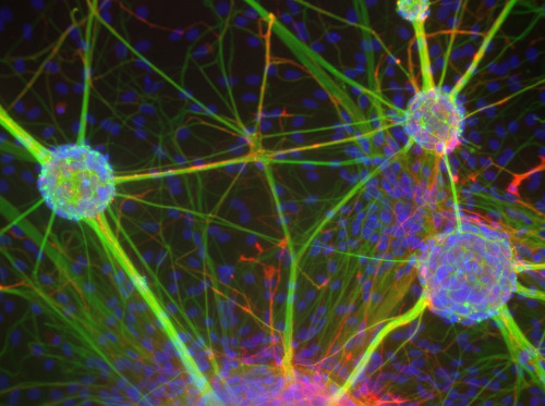
The birth of new neurons (neurogenesis) does not end completely during development but continues throughout all life in two areas of the adult nervous system, i.e. subventricular zone and hippocampus. Recent research has shown that hippocampal neurogenesis is crucial for memory formation. These studies, however, have not yet clarified how the newborn neurons are integrated in the existing circuits and thus contribute to new memories formation and to the maintenance of old ones.
The team of researchers of CNR-LUMSA-EBRI at the European Centre for Brain Research, organization established in Rome with the key contribution of the Santa Lucia Foundation, has taken a step forward to understand the requirements of newborn neurons in the process of learning and memory. The neuroscientists coordinated by dr. Felice Tirone of the Institute of Neurobiology and Molecular Medicine (INMM) of CNR, in collaboration with prof. Vincenzo Cestari of the Institute for Neuroscience of CNR and the LUMSA University and with dr. Alberto Bacci of the European Brain Research Institute, have shown that a key factor for neurogenesis is represented by the speed of differentiation of progenitors (stem cells that give rise to neurons) in hippocampus. From such speed will in fact depend the success of the whole process. “New neurons must maturate according to a correct temporal sequence in order to become able to acquire new memories and retrieve the existing ones”, explains Tirone.
This study is based on a new experimental approach, that involves the generation of a mice line in which the differentiation of newborn neurons is accelerated without altering their number. This is obtained by the selective expression in neural progenitors of the hippocampus of PC3/Tis21, a gene specifically able to accelerate the differentiation of these and other types of neural progenitors.
“Compelling new neurons to rush ahead in their differentiation for a predefined time period, we have observed that a small number of neurons of 2-3 weeks of age is critical for learning,” continues the INMM-CNR researcher. “In fact, mice so treated are not only unable to learn new spatial information, but they are also unable to use previously acquired memories.”
“PC3/Tis21, of which we have previously observed an action against brain tumors in consequence of its ability to promote differentiation of neural progenitor cells, might indeed have other practical outcomes” continues Tirone “since it is activated by Nerve Growth Factor, a molecule whose deprivation appears to be an important component of Alzheimer’s disease. In fact hippocampus is one of the first brain regions damaged by Alzheimer’s disease, which is characterized mainly by temporal and spatial disorientation and by a memory deficit. We might gain some useful information also to understand the mechanisms underlying this disease.”
Thus, it is an open and relevant question in neuroscience the understanding of mechanisms and factors that control or influence adult neurogenesis, in the field of memory as these researches now indicate, but also of depression, which, as some researchers have suggested, might take place in consequence of a defective adult neurogenesis.
Article: Farioli-Vecchioli S, Saraulli D, Costanzi M, Pacioni S, Cinà I, et al. (2008) The Timing of Differentiation of Adult Hippocampal Neurons Is Crucial for Spatial Memory. PLoS Biol 6(10): e246
27.10.2008
Немного о нейрогенезе
18.10.2008
Допаминовые рецепторы и контоль веса.
Вот они какие - мои любимые... :))) Нашла инфу на http://www.scientificblogging.com/news_releases/dopamine_signaling_why_some_like_chocolate_milkshakes_too_much
Dopamine Signaling - Why Some Like Chocolate Milkshakes Too Much
Submitted by News Staff on 16 October 2008 - 2:00am. Neuroscience
Using brain imaging and chocolate milkshakes, scientists have found that women with weakened "reward circuitry" in their brains are at increased risk of weight gain over time and potential obesity. The risk increases even more for women who also have a gene associated with compromised dopamine signaling in the brain.
The results, drawn from two studies using functional magnetic resonance imaging (fMRI) at the University of Oregon's Lewis Center for Neuroimaging, appear in the Oct. 17 issue of the journal Science. The first-of-its-kind approach unveiled blunted activation in the brain's dorsal stratium when subjects were given milkshakes, which may reflect less-than-normal dopamine output.
"Although recent findings suggested that obese individuals may experience less pleasure when eating, and therefore eat more to compensate, this is the first prospective evidence for this relationship," said Eric Stice, lead author and senior researcher at the Oregon Research Institute (ORI) in Eugene. "The evidence of temporal precedence suggests it is a true vulnerability factor that predates obesity onset. In addition, the evidence that this relation is even stronger for individuals at genetic risk for compromised signaling in these brain regions points to an important biological factor that appears to increase risk for obesity onset."
Stice, who has a courtesy appointment in the UO psychology department, has studied eating disorders and obesity for 18 years. He was joined in the new research by ORI colleague Sonja T.P. Spoor, Cara Bohon, a UO doctoral student in clinical psychology, and Dana M. Small of the John B. Pierce Laboratory in New Haven, Conn., and the Yale University School of Medicine.
Dopamine is the primary neurotransmitter in the brain's reward pathways. Food intake is associated with dopamine release, while pleasure from eating correlates with the amount of dopamine release. Previous studies have suggested that obese individuals have fewer dopamine receptors in the brain and have to eat more than lean people to be satisfied.
Using fMRI, Stice's team measured the extent to which the dorsal striatum was activated in response to an individual's receipt of a taste of chocolate milkshake or a tasteless solution.
One study involved 43 female college students ranging in age from 18 to 22 with a mean body mass index (BMI) of 28.6. The second study looked at 33 adolescent girls, ages 14-18, with a mean BMI of 24.3. Most of the participants were tested for the presence of a genetic variation known as the Taq1A1 allele, which is linked to a lower number of dopamine D2 receptors.
Researchers tracked changes in BMI over a year. Results showed that participants with decreased striatal activation in response to receiving milkshake and those with the A1 allele were more likely to gain weight over time.
"I was quite excited by the study itself, as it is the first prospective study to utilize fMRI and genetic data to predict unhealthy weight gain," said Bohon, whose doctoral work is done on both the UO campus and the nearby ORI facility. "The findings suggest that certain biological factors may impact one's risk for weight gain, which is important in order to better understand how we can eventually intervene and prevent obesity."
10.10.2008
Морфий больше не актулен!
Встречайте новооткрытый белочек! Прочитала на http://www.biologynews.net/archives/2008/10/08/cell_protein_suppresses_pain_8_times_more_effectively_than_morphine.html
More people suffer from pain than from heart disease, diabetes and cancer combined, but many of the drugs used to relieve suffering are not completely effective or have harmful side effects.
Now researchers at the University of North Carolina at Chapel Hill School of Medicine and the University of Helsinki have discovered a new therapeutic target for pain control, one that appears to be eight times more effective at suppressing pain than morphine.
The scientists pinpointed the identity and role of a particular protein that acts in pain-sensing neurons, or nerve cells, to convert the chemical messengers that cause pain into ones that suppress it.
"This protein has the potential to be a groundbreaking treatment for pain and has previously not been studied in pain-sensing neurons," said lead study author Mark J. Zylka, Ph.D., assistant professor of cell and molecular physiology at UNC. The results of the study will be published online in the journal Neuron, on Wednesday (Oct. 8) and in the print edition the following day.
The biological basis of pain is complex. To study the transmission of painful signals throughout the body, many researchers use "marker" proteins that label pain-sensing neurons. One such marker, FRAP (fluoride-resistant acid phosphatase), has been employed for this purpose for nearly 50 years, but the gene that codes for its production was never identified.
That is, until researchers at UNC found that FRAP is identical to PAP (prostatic acid phosphatase), a protein routinely used to diagnose prostate cancer whose levels increase in the blood of patients with metastatic prostate cancer.
Previous research hinted that FRAP and PAP may have a shared identity. To determine whether or not this was the case, Zylka teamed up with Dr. Pirkko Vihko, a professor from the University of Helsinki who had genetically engineered mice that were missing the gene for PAP. When Zylka and his colleagues studied tissues from these mutant mice, they were happy to see that FRAP activity was missing. This revealed that the two proteins were in fact identical.
Further, the mutant mice proved more sensitive than normal mice to inflammatory pain and neuropathic pain, two common forms of chronic pain in humans. These increased sensitivities diminished when researchers injected excess amounts of PAP into the spinal cords of the mutant mice.
"We were really blown away that a simple injection could have such a potent effect on pain," Zylka said. "Not only that, but it appeared to work much better than the commonly used drug morphine."
The new protein suppressed pain as effectively as morphine but for substantially longer. One dose of PAP lasted for up to three days, much longer than the five hours gained with a single dose of morphine.
The next question for the researchers was how PAP suppressed pain. It is already known that when pain-sensing neurons are stimulated, they release chemicals known as nucleotides, specifically adenosine triphosphate (ATP). This in turn sets off the events that invoke a painful sensation. But if ATP degrades to adenosine, that inhibits the neurons that transmit pain signals, thus relieving pain. Through a series of experiments, the UNC researchers showed that PAP removes the phosphate group, generating adenosine. Their study is the first to identify and characterize the role of such a protein in pain-sensing neurons.
Zylka and his colleagues are now searching for additional proteins that degrade nucleotides in these neurons. They are also working to develop small molecules that interact with PAP to enhance or mimic its activity.
"It is entirely possible that PAP itself could be used as a treatment for pain, through an injection just like morphine," Zylka said. "But we would like to modify it to be taken in pill form. By taking this field in a new direction, we are encouraged and hopeful that we will be able to devise new treatments for pain."
Source : University of North Carolina School of Medicine


