


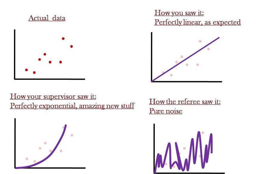
Вот так всё обычно и происходит...

Не болейте! :)


Calorie Restriction May Make Rats Live Longer, But Doesn’t Work For PrimatesСпасибо за наводку jtotheizzoe.
Remember those famous studies in worms, mice and rats that indicated that a restricted-calorie diet increased lifespan? Well, the results of a 30-year study in rhesus monkeys has just been reported, and the results make it pretty clear that it doesn’t apply to primates … likely including us.
The molecular pathways and gene circuits that are activated in our bodies as a result of long-term calorie restriction are immensely complicated. Like a tangled ball of yarn complicated. We don’t understand how they work, frankly, and there’s just no magic bullet when it comes to diet and lifespan in creatures like monkeys and humans (I’m looking at you, resveratrol). It’s certainly good for one’s long-term survival not to overeat into Honey-Boo-Boobesity, but what does current science say about calories, long life and naked apes?
Studies of humans who live past 100 point to genetics as more important than restricting calories. Which genes? Time may tell. And none of this has helped us understand how different diet compositions may affect aging.
Read more at Nature News.

Информация о возможных способах лечения болезни Паркинсона стволовыми клетками описан на http://www.eurekalert.org/pub_releases/2010-05/nioc-esc050610.php
Читаю на http://www.biologynews.net/archives/2010/04/12/mit_new_cell_measurement_system.html
Using a sensor that weighs cells with unprecedented precision, MIT and Harvard researchers have measured the rate at which single cells accumulate mass — a feat that could shed light on how cells control their growth and why those controls fail in cancer cells.
The research team, led by Scott Manalis, MIT associate professor of biological engineering, revealed that individual cells vary greatly in their growth rates, and also found evidence that cells grow exponentially (meaning they grow faster as they become larger).
The new measurement system, reported in the April 11 edition of the journal Nature Methods, is the first technique that can measure cells' mass as they grow over a period of time, ranging from five to 30 minutes. Previous methods for measuring cell growth rates have focused on volume or length measurements, and have not yet exhibited the necessary precision for revealing single cell growth models.
How they did it: The cell-mass sensor, which Manalis first demonstrated in 2007, consists of a fluid-filled microchannel etched in a tiny silicon slab that vibrates inside a vacuum. As cells flow through the channel, one at a time, their mass slightly alters the slab's vibration frequency. The mass of the cell can be calculated from that change in frequency, with a resolution as low as a femtogram (10-15 grams).
Michel Godin, a former postdoctoral associate in Manalis' lab and co-lead author of the paper, developed a way to trap a cell within the microchannel by precisely coordinating the flow direction. That enables the researchers to repeatedly pass a single cell through the channel every second or so, measuring it each time it moves through.
The researchers studied four types of cells: two strains of bacteria (E. coli and B. subtilis), a strain of yeast and mammalian lymphoblasts (precursors to white blood cells). They showed that B. subtilis cells appear to grow exponentially, but they did not obtain conclusive evidence for E. coli. That's because there is so much variation between individual cell growth rates in E. coli, even for cells of similar mass, says Francisco Delgado, a grad student in Manalis' lab and co-lead author of the paper.
If cells do grow exponentially, it means there must be some kind of mechanism to control that growth. Otherwise, when cells divide into two slightly different-sized daughter cells, as they often do, the larger cell in each generation would always grow faster than the smaller cell, leading to inconsistent cell sizes.
"If there were no control over the process, the variation in cell size would be all over the map," says Marc Kirschner, professor of systems biology at Harvard Medical School and an author of the paper. However, biologists don't know yet how that control mechanism might work.
Next steps: In their current studies, the researchers are tagging proteins inside the cell with fluorescent molecules that reveal what stage of the cell cycle the cell is in, allowing them to correlate cell size with cell cycle position. They are also working on a way to add chemicals such as nutrients, antibiotics and cancer drugs to the fluid inside the microchannel, so they can study how those substances affect growth rates.
Source : Massachusetts Institute of Technology
Две мои любимейшие темы в одном чудесном посте. Автор - Ed Yong
All of our cells are staffed by armies of executioners. They are usually restrained but when unleashed, they can set off a fatal chain reaction that kills the cell. This suicide squad does away with billions of cells every day. It helps to balance the production of new cells with the loss of old ones, to sculpt growing tissues and to destroy potential cancer cells.
But a new study suggests that the executioners aren't always lethal. In fact, they're essential for life. Through the unorthodox method of damaging our DNA, they can actually activate important genes. This technique for switching genes on is new to science but it's apparently vital for allowing some types of stem cell to produce new types of tissue.
Stem cells are bundles of untapped potential, with the ability to produce hundreds of specialist cells across the body. This process is called differentiation. Its details vary depending on which type of cell is being produced, but scientists have recently found that some aspects are apparently common to all tissues, be they muscle, blood or bone. Surprisingly, one of these is the recruitment of executioner proteins - caspases.
Caspases cut up other proteins and in doing so, some of them produce yet more caspases. The result is a growing army of death, hacking and slashing its way through the cell. But one of these killers - caspase-3 - is a necessary part of differentiation. Get rid of it and, suddenly, stem cells can't produce their specialised daughters. Now, thanks to Brian Larsen from the Sprott Centre for Stem Cell Research, we know why.
Caspase-3 activates a protein called CAD (or caspase-activated DNase in full) by slicing apart other proteins holding it at bay. Once released, CAD lives up to its villainous acronym. It pairs up with an identical twin to create a molecule that looks and acts like a pair of scissors. The pair can cut DNA, cleaving the famous double helix in two. These sorts of cuts are normally very bad news for a cell. If they aren't repaired quickly and accurately, the consequences can include death or cancer.
But Larsen has found that stem cells deliberately break their own DNA by recruiting caspase-3 and CAD. This act of self-harm switches on important genes that are needed for differentiation; without it, the generalist cells can't specialise. This is an entirely new way of activating genes and it appears to be both important and widespread.
Larsen studied stem-like cells called myoblasts, which give rise to various types of muscle cells. As the myoblasts differentiated, Larsen watched for signs of shattered DNA using a clever test called the 'comet assay'. The technique involves puncturing a cell and placing it in an electric field. The field drives DNA through the punctured cell but only if it has already been broken into small pieces. If it has, it appears as a streak outside the cell, rather like the tail of a comet.
Sure enough, the comet test revealed that differentiating cells suffer from significant amounts of damaged DNA. Thankfully, the injuries are only temporary and the cells soon marshal their repairmen to fix the breaks.
The myoblasts need these breaks to produce muscle fibres and to create the breaks, they rely on caspase-3 and its ability to activate CAD. Larsen managed to block the development of muscle fibres by dousing myoblasts with chemicals that neutralise caspase-3. The same thing happened if he used cells with mutant versions of CAD, which couldn't be activated. In both cases, the cells failed to show any signs of broken DNA.
CAD targets a gene called p21 that's absolutely necessary for the development of muscle and plenty of other tissues. Larsen found that CAD cuts p21's 'promoter', a stretch of DNA lying next to the gene that's responsible for switching it on. Somehow, these cuts activate the gene. It's still not clear how this works, but Larsen has some ideas. The cuts could change how the surrounding DNA is packaged, exposing the p21 gene and making it easier to 'read'. Alternatively, the cuts could remove chemical 'marks' attached to the DNA that would otherwise silence it.
Damaging your own DNA may seem like a rather extreme tactic for a cell to take but it's not unheard of as a deliberate ploy. Whenever we face new infections, our body generates antibodies by breaking the DNA of special genes, stitching them back together in new combinations. That's a very controlled process, but so is the damage that leads to differentiation. It's a careful surgical strike, rather than a shock and awe campaign.
During differentiation, Larsen found that DNA breaks are actually few and far between. They appear to be carefully orchestrated so that the entire genome doesn't become a shattered mess. This precision is even more remarkable when you consider that CAD cuts DNA indiscriminately, with little care for specific sequences. Larsen thinks that CAD is constrained by the way the DNA is packaged, so that only places that are meant to be cut are exposed for slicing and dicing. Only further experiments will tell if he is right.
Reference: Larsen et al. 2010. Caspase 3/caspase-activated DNase promote cell differentiation by inducing DNA strand breaks. http://dx.doi.org/10.1073/pnas.0913089107
Статья с http://scienceblogs.com/notrocketscience/2010/02/our_cells_produce_new_tissues_by_recruiting_executioners_to.php
Какая прелесть! Это ведь именно то, что нужно сейчас нашим культуральщикам! Трёхмерная культура клеток!!! Это, конечно, не 22 и даже не 11 измерений, но нет ничего невозможного... Информация с http://www.biology-blog.com/blogs/permalinks/9-2008/reversible-3-d-cell-culture-gel-invented.html
Reversible 3-D cell culture gel invented
Reversible 3-D cell culture gel invented
Singapore's Institute of Bioengineering and Nanotechnology (IBN), which celebrates its fifth anniversary this year, has invented a unique user-friendly gel that can liquefy on demand, with the potential to revolutionize three-dimensional (3D) cell culture for medical research.
As reported in Nature Nanotechnology (Y.S. Pek, A. C. A. Wan, A. Shekaran, L. Zhuo and J. Y. Ying, "A Thixotropic Nanocomposite Gel for Three-Dimensional Cell Culture"), IBN's novel gel media has the unique ability to liquefy when it is subjected to a moderate shear force and rapidly resolidifies into a gel within one minute upon removal of the force. This phenomenon of reverting between a gel and a liquid state is known as thixotropy.
IBN's thixotropic gel is synthesized from a nanocomposite of silica and polyethylene glycol (PEG) under room temperature, without special storage conditions. This novel material facilitates the safe and convenient culture of cells in 3D since cells can be easily added to the gel matrix without any chemical processes.
As per IBN Executive Director Jackie Y. Ying, Ph.D., "Cell culture is conventionally performed on a flat surface such as glass slides. It is an essential process in biological and medical research, and is widely used to process cells, synthesize biologics and develop therapys for a large variety of diseases.
"Cell culture within a 3D matrix would better mimic the actual conditions in the body as in comparison to the conventional 2D cell culture on flat surfaces. 3D cell culture also promises the development of better cell assays for drug screening," Dr. Ying added.
Another key feature of IBN's gel is the ease with which scientists can transfer the cultured cells from the matrix by pipetting the mandatory amount from the liquefied gel.
Unlike conventional cell culture, trypsin is not mandatory to detach the cultured cells from the solid media. As trypsin is an enzyme that is known to damage cells, particularly in stem cell.
cultures, the long-term quality and viability of cells cultured using IBN's thixotropic gel would improve substantially without the exposure to this enzyme.
Scientists are also able to control the gel's stiffness, thus facilitating the differentiation of stem cells into specific cell types.
"Ways to control stem cell differentiation are important as stem cells can be differentiated into various cell types. Our gel can provide a novel method of studying stem cell differentiation, as well as an effective new means of introducing biological signals to cells to investigate their effect in 3D cultures," said Shona Pek, IBN Research Officer.
Andrew Wan, Ph.D., IBN Team Leader and Principal Research Scientist, added, "Another interesting property of the gel is its ability to support the extracellular matrix (ECM) secretions of cells. Gel stiffness is modulated by ECM secretions, and can be used to study ECM production by cells responding to drug therapys or disease conditions.
"The thixotropic gel may then provide new insights for basic research and drug development," Dr. Wan added.
Posted by: Janet http://www.a-star.edu.sg/astar/index.jsp
Мама крови. Вот она! Наконец, эмбриональная предшественница клеток крови - чётко определена. Информация с http://www.scientificblogging.com/news_releases/discovery_mother_of_all_blood_stem_cells
Discovery - 'Mother' Of All Blood Stem Cells
Submitted by News Staff on 29 August 2008 - 1:00am. Developmental
Johns Hopkins researchers say they have discovered the earliest form of human blood stem cells and deciphered the mechanism by which these embryonic stem cells replicate and grow. They also found a surprising biological marker that pinpoints these stem cells, which serve as the progenitors for red blood cells and lymphocytes.
The research reported today used federally approved embryonic stem cell lines.
The biochemical marker, angiotensin-converting enzyme (ACE), is well known for its role in the regulation of blood pressure, blood vessel growth, and inflammation. ACE inhibitors are already widely used to treat hypertension and congestive heart failure, and the findings are, the researchers say, likely to hold promise for developing new treatments for heart diseases, anemias, leukemia and other blood cancers, and autoimmune diseases because they show for the first time that ACE plays a fundamental role in the very early growth and development of human blood cells.
"We figured out how to get the 'mother' of all blood stem cells with the right culture conditions," says Elias Zambidis, M.D., Ph.D., of the Institute of Cell Engineering at the Johns Hopkins University School of Medicine and the Division of Pediatric Oncology at the Sidney Kimmel Comprehensive Cancer Center at Johns Hopkins.
"There is real hope that in the future we can grow billions of blood cells at will to treat blood-related disorders, and just as critically if not more so, we've got ACE as a 'new' old marker to guide our work," Zambidis adds.
Researchers did not expect ACE to have a role in blood stem cells, he notes, "but were very pleasantly surprised to discover it as a beacon for finding the earliest blood stem cells known, as well as new ways to find and manipulate this marker to make them grow."
The team's findings, published Aug. 26 in the online edition of the journal Blood, explain that these earliest stem cells marked by ACE, called hemangioblasts, first arise normally in the developing human fetus, when a woman is three or four weeks pregnant. Hemangioblasts can now be derived in unlimited supply experimentally from cultured human embryonic stem cells, which are the origin of all cell types in the body. These hemangioblasts go on to become either blood cells or endothelial cells, which form the inner lining of the heart, veins and arteries, and lymph vessels.
The research grew out of Zambidis' interest in understanding the complex biological processes of blood development and the transformation of embryonic stem cells into the various types of cells that make up the human body.
Hemangioblasts make the body's earliest form of blood in the fetal yolk sac, which nourishes a fertilized egg, and later in the fetal liver and bone marrow. However, because human embryonic cells disappear early in gestation, their role in the early production of blood could not, to the researchers' knowledge, be studied in humans because scientists had no way to identify these human progenitor blood stems cells to follow their development. The scientists suspected they existed in humans, however, because they have been found in mice and zebra fish.
To find the blood stem cell, Zambidis' team grew human embryonic stem cells in culture and fed them growth factors over 20 days. Each time the cell colonies expanded, the researchers sampled individual cells, searching for ones capable of making both endothelial and blood cells, the hallmark of hemangioblasts.
They plucked the newly discovered hemangioblasts from culture dishes, grew them in conditions that Zambidis and his team developed to speed replication, and tested cells for their ability to make endothelial and blood cells. Cells capable of making endothelial cells and all the elements of blood (platelets, and white and red cells) were specifically marked with ACE on their outer surface.
The researchers found not only that ACE was a marker for hemangioblasts, but turning off the enzyme also helps guide the cells' replication and maturation into either blood or endothelial cells. By treating the hemangioblasts with losartan, an ACE pathway blocking agent routinely used to treat high blood pressure, dramatically increased the rate of blood cell production.
The next step, Zambidis adds, is to test this research in animal models and show that "we can make lots and lots of blood cells from human stem cells for transfusions, regenerate new vascular trees for heart diseases, as well as create test tube factories for making transplantable blood cells that treat diseases. We are very far from treatment," Zambidis cautions, "but this is a big step."
If the new technique of mass producing progenitor blood cells is eventually proven to work in humans, it would allow patients getting bone marrow transplants to have their own stem cells creating the blood they need, significantly reducing rejection risk.
The research reported today used federally approved embryonic stem cell lines, but other related research by the team comes from nonapproved lines. The study was supported by grants from the National Institutes of Health and the Maryland Stem Cell Research Fund.
Ну разве не радость? Прочитала на http://www.scientificblogging.com/news_releases/50_year_cell_division_mystery_solved_say_researchers
Researchers from Oregon State University say they have resolved a controversy that cellular biologists have been arguing over for nearly 50 years, with findings that may aid research on everything from birth defects and genetic diseases to the most classic "cell division" issue of them all – cancer.
The exact mechanism that controls how chromosomes in a cell replicate and then divide into two cells, a process fundamental to life, has never been completely pinned down, researchers say. You can find the basics in any high school biology textbook, but the devil is in the details.
"Researchers have been debating cell cleavage ever since the cell was discovered, with two basic models proposed around 1960 of how a contractile ring pulls together and allows a single cell to split into two," said Dahong Zhang, an OSU associate professor of zoology. "Part of the problem is that until now there was no decisive way to manipulate the cytoskeleton, such as the microtubules and filaments that are involved, and see what was happening as it occurred."
To address that, Zhang developed some new instrumentation that uses "microneedles" and state-of-the-art imaging techniques which allow direct manipulation of the cytoskeleton, while capturing the results of contractile ring formation. The system has not only solved this decades-old riddle, but "the technology is a very powerful new approach," Zhang said, that should find applications in other cell biology research issues.
It has been known for some time, scientists say, that a "contractile ring," which is composed of some of the same fibers used in muscle contraction, move into the correct position, pull and split a cell in two after its chromosomes have been separated. This is distribution of genetic materials at its most basic level, and it has to be done at exactly the right place and time. When the process breaks down, cancer and other serious medical or genetic issues can be a result.
But if you think of the cell as a sphere, what was less clear was whether the "equator" contracted or the "poles" relaxed to allow this contraction and division. Two distinct theories were formed, called polar relaxation and equatorial stimulation, to explain this aspect of cell division – and some scientists have spent much of their careers arguing for one side or the other.
Turns out, Zhang said, that both sides were correct. Nature and evolution have actually created a basic way for a cell to divide with a backup system that can work if the other approach fails.
"Accurate cell division is one of the most critical of all life functions, and there clearly is an evolutionary value to having redundancy, a system able to do it two different ways," Zhang said. "It makes perfect sense when you think about it. The findings speak plainly for themselves, and there should no longer be a question over which model is right."
By labeling cells and moving microtubules around while still being able to see them and their impact on microfilaments, OSU researchers were able to selectively inhibit one mechanism of cell division or the other. They discovered that in the same cell type, it could divide either by polar relaxation or equatorial stimulation – the two mechanisms are not mutually exclusive.
The findings, Zhang said, add significantly to the basic understanding of cell biology, and should be of special interest to cancer researchers. Cancer is essentially the loss of normal control over cell division and migration. In fact, a compound used in Zhang's laboratory to inhibit cell division while they studied it was taxol – a commonly used cancer drug.
Accurate and effective cell division, researchers say, is also key to the understanding of some genetic diseases, miscarriages, birth defects and other issues.
The studies were supported by the National Science Foundation and the American Heart Association.


